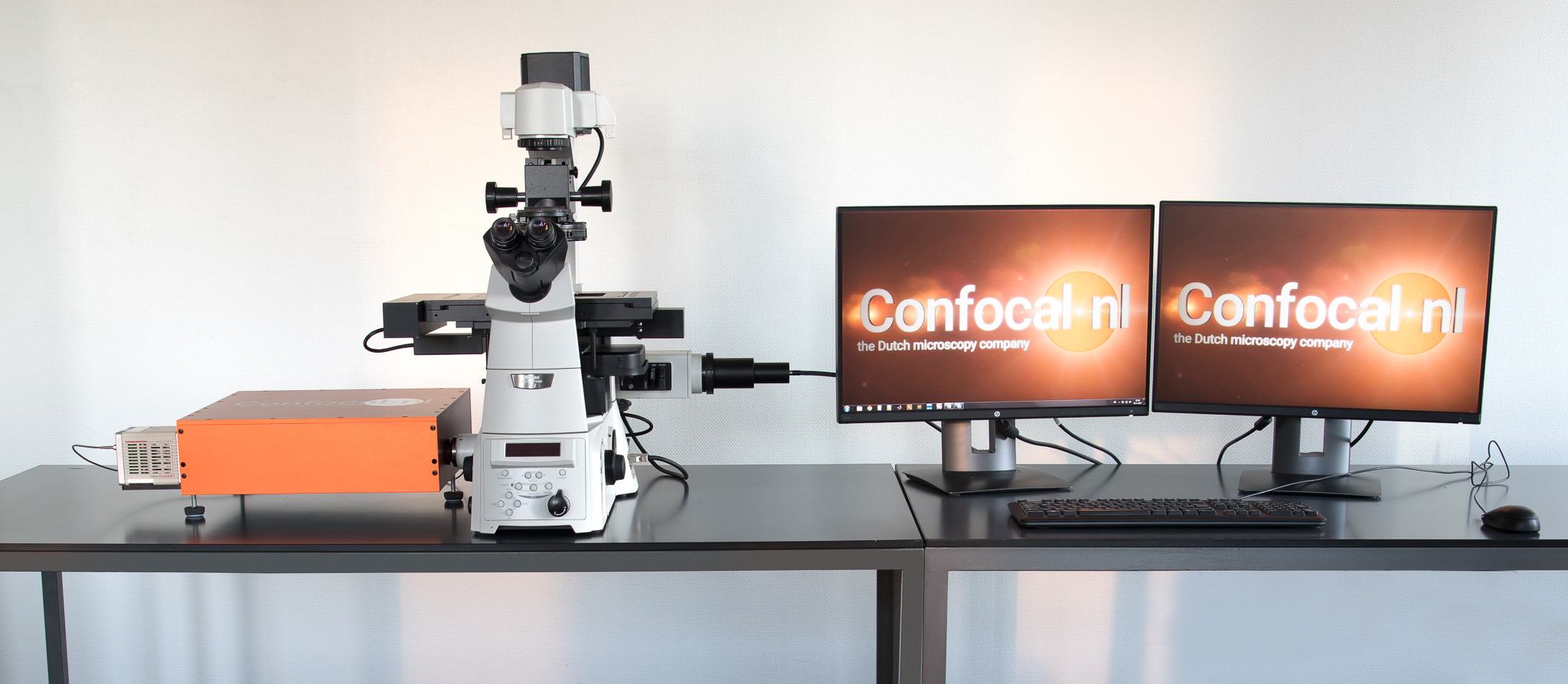
Rescan Confocal Microscope

The system architecture of the RCM provides a standard C-mount interface with microscope and camera. The RCM includes two scanning mirror sets: the scanner and the re-scanner. While the scanner scans the specimen, the re-scanner writes the emission signal on the camera chip. By doubling the sweep of the re-scanner, the RCM image has an increased resolution. Because the RCM is based on opto-mechanics only, no post processing is needed to achieve the high resolution images. As described in "Re-scan confocal microscopy: scanning twice for better resolution; G. M. R. De Luca, R. M. P. Breedijk, E. M. M. Manders et al. Biomedical Optics Express 4,2644-2656 (2013)", this improved resolution is pinhole independent and therefore a strongly enhanced photon-efficiency (and lower photo-toxicity) is observed. Additional deeper confocality in the sample is established.
In the RCM unit, the excitation path is similar as traditional confocal systems (the blue light path in the image above).The unique feature of the RCM is based on the additional re-scanning mirrors used for the emission fluorescence (the green lightpath above), which is written on a sensitive camera chip. Together with the open pinhole (AU=2), this provides a exceptional image with high resolution and superior signal to noise ratio. The lateral resolution of the RCM is increased to 170nm compared to 240nm of regular confocal microscope and the signal to noise ratio is 4x
better.
Standard Features
- Lasers: Support up to 4 laser lines: 405, 488, 568, and 638nm , Additional laser wavelengths upon request, 3rd party laser combiner supported, Laser output through single line fiber (FC Connector)
- Microscope: Standard microscope with C-mount adapter , Major brand microscopes supported
- Camera: Hamamatsu ORCA-Flash 4.0 , Other major EMCCD/sCMOS camera supported
- Features: 170nm lateral resolution , 600nm axial resolution , 1fps (@ 512 x 512), 50um (2.2 AU for 100x objective), Optimized for 100x, 60x and 40x objectives,
- Software: Image acquisition software included , Other major image processing software packages supported


